
Abstract
Lower facial enhancement represents one of the most technically challenging interventions in aesthetic medicine. The jawline and chin, as critical skeletal boundaries, define lower facial structure, balance the profile, and carry implications for both youth and gender aesthetics. Unlike volumisation of the midface or periorbital region, successful lower face augmentation requires a skeletal mindset: linear precision, spatial control, and anatomical discipline. Central to this endeavour is visual planning—especially when performed using sterile, high-fidelity tools such as the SKRYBE FMD marker. This article explores the anatomical rationale, mapping methodology, and safety protocols underpinning effective chin and jawline augmentation, with an emphasis on mapping as a procedural scaffold.
Introduction
In facial aesthetics, there are few regions as simultaneously rewarding and unforgiving as the lower third of the face. The mandible serves not only as a physical boundary for facial proportions, but also as an expressive platform: shaping the interplay between strength, youth, and symmetry. With the rise of non-surgical techniques, the ability to reshape this zone using injectable fillers has advanced significantly. Yet, precisely because the lower face is bounded by skeletal landmarks and defined angles, even minor deviations in technique can produce disproportionate aesthetic consequences.
To approach this area without comprehensive pre-injection planning is to invite inconsistency. The cornerstone of modern treatment in this zone is strategic, visible mapping. When executed correctly, visual markings guide not only where to inject, but how—and why.
Mandibular Aging and Structural Regression
The aging mandible is not simply a platform upon which changes are seen; it is itself subject to profound structural regression. Decades of cephalometric and cadaveric research have established predictable patterns of change with advancing age. These include retraction of the pogonion (the most anterior point of the chin), descent of the menton (the lowest midline point of the mandible), and progressive blunting of the gonial angle【1】.
In clinical terms, this translates to a downward and posterior drift in chin projection, elongation of the lower third, and loss of mandibular continuity at the pre-jowl sulcus. These changes displace the perceived centre of gravity of the face, altering its balance and softening cues of strength and youth. The role of the practitioner, therefore, is not simply to add volume, but to re-establish skeletal proportion with anatomical fidelity.
The Value of Visual Mapping
Pre-injection visual mapping serves multiple clinical purposes. It translates a three-dimensional structural assessment into a consistent two-dimensional surface guide. It enables symmetry checks in real time, supports cannula trajectory control, and assists in documentation. Most importantly, it anchors the procedure within defined anatomical zones, reducing variability and risk.
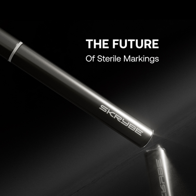
The use of sterile, high-contrast white markers, such as the SKRYBE FMD, allows these markings to persist through antiseptic cleansing and during the dynamic manipulation of tissue throughout the procedure. This ensures that once the skin is prepped, the anatomical plan remains both visible and sterile. Marking before cleansing, or attempting to re-mark after, is discouraged as it introduces both alignment drift and infection risk.

Chin Augmentation: Principles and Planning
The chin is the visual fulcrum of the lower face. Its projection, width, and vertical height determine not only individual attractiveness but also gender characterisation and profile balance. In aesthetic correction, the goal is rarely simple enlargement; rather, it is precise structural calibration.
Mapping begins with the establishment of a midline, typically using the philtrum and labiomental crease as reference points. The current pogonion is then identified in profile, and the desired projection marked. Lateral tubercles, which define chin width and contour, are marked approximately 1.5 cm from midline in most adults. The menton, located at the chin’s inferior edge, helps determine whether augmentation will elongate or simply advance the lower third.
Injections are typically placed at the supraperiosteal level using 22–25G cannulas. Product volumes range from 0.5 to 1.2 mL depending on existing projection and soft tissue coverage. Care is taken to avoid inferior elongation of the chin in patients where lower third height is already generous.
Mapping confers significant advantages: it maintains midline control, prevents overfilling of the central chin, and allows the injector to assess symmetry in multiple vectors prior to committing to filler placement.
Addressing the Pre-Jowl Sulcus
The pre-jowl sulcus is formed by a combination of mandibular resorption and soft tissue descent, creating a concavity anterior to the jowl proper. If uncorrected, it breaks the visual continuity of the mandibular border. Yet, overcorrection—particularly when performed without mapping—risks creating unnatural bulk or inadvertently filling the jowl itself.
Surface planning involves drawing the mandibular border from pogonion to gonial angle, followed by identification of the jowl apex. A shallow crescent anterior to the jowl is then marked, outlining the sulcus zone.
Treatment is typically performed in the supraperiosteal or deep subcutaneous plane using a fanning technique. A volume of 0.3 to 0.8 mL per side is usually sufficient. Marking enables the injector to preserve mandibular angulation and avoid disrupting adjacent contour.
Redefining the Mandibular Border
A clean, continuous mandibular line is a hallmark of youthful structure. Age-related regression blurs this contour, especially in the lateral two-thirds. Mapping the border using a linear marking from menton to gonial angle allows the practitioner to assess horizontal symmetry and plan filler placement in consistent, reproducible intervals.
Injections along the mandibular body are best performed supraperiosteally or sub-SMAS, depending on the patient's soft tissue thickness. Cannula use is advised to minimise trauma and permit linear filler threads. Volume typically ranges from 0.5 to 1.0 mL per side.
Visual planning mitigates common aesthetic errors, including overcorrection, “step-off” deformities, and lateral asymmetry. Moreover, it provides a visible scaffold to evaluate how the projected line aligns with the patient's natural mandibular angulation.
Gonial Angle Definition
The gonial angle—formed by the intersection of the posterior mandibular body and the ascending ramus—serves as a critical determinant of facial width and jawline strength. Its prominence varies by gender and must be treated with restraint, especially in female patients.
Mapping requires palpation of the angle during clenching, followed by a mark placed approximately 1 cm above the bony landmark to prevent inferior migration of filler. Treatment is delivered supraperiosteally with 0.3 to 0.7 mL per side. Without visual reference, there is increased risk of asymmetry, overprojection, or placement within the masseter muscle, which may distort functional or aesthetic outcomes【2】.
Anatomical Safety Considerations
All markings must also account for anatomical caution zones. The mental foramen, typically located below the second premolar, should be avoided within a 1.5 cm radius. Similarly, the facial artery and vein, which curve anterior to the mandibular border, must be identified and avoided through mapping and, where applicable, cannula orientation【3【4】.
Sterile white-ink markings ensure these structures remain visible post-preparation, reducing the cognitive burden during injection and facilitating safe navigation in anatomically variable patients.
Clinical Integration and Documentation
Visual mapping should be conducted immediately after antiseptic skin preparation. The use of a sterile SKRYBE FMD marker ensures that all landmarks—midline, sulci, tubercles, angles—remain present throughout the procedure.
Photographic documentation of the marked face provides medico-legal reassurance, allows comparative follow-up, and supports reproducibility across sessions. The visual plan becomes not only a guide for injection but also a teaching and audit tool.
Conclusion
The use of sterile, high-contrast markers—such as SKRYBE—represents the modern standard for injector precision, safety, and professionalism. When planning is made visible, treatment becomes reproducible. And when mapping becomes habitual, results become dependable.
References
- Mendelson B, Wong CH. Changes in the facial skeleton with aging: implications and clinical applications in facial rejuvenation. Aesthetic Plast Surg. 2012;36(4):753–760. https://doi.org/10.1007/s00266-012-9864-z
- Ahmed F, Massry GG. Complications of lower face filler: recognizing masseteric hypertrophy, overcorrection and gonial distortion. J Cosmet Dermatol. 2021;20(1):13–18. https://doi.org/10.1111/jocd.13479
- Hu KS, Kwak HH, Song WC, Koh KS, Kim HJ. Branching patterns of the mental nerve and variability of the mental foramen. J Craniofac Surg. 2007;18(6):1325–1329. https://doi.org/10.1097/scs.0b013e318159ec25
- Koziej M, Polak J, Wysocki J, et al. Facial artery: a comprehensive review of its anatomy, variations and clinical significance. Ann Anat. 2021;233:151611. https://doi.org/10.1016/j.aanat.2020.151611


