
Abstract
The labiomental fold, often overlooked in standard lower face assessments, represents a critical transitional landmark between the lower lip and the chin. Its exaggeration with age contributes significantly to the appearance of perioral ageing, profile imbalance, and dynamic facial disharmony. While filler-based rejuvenation of this region has become increasingly common, poor anatomical understanding and suboptimal planning frequently result in either under-correction or unnatural distortion. This article examines the layered anatomy of the labiomental complex, common morphological variants, and the role of visual mapping—particularly with sterile white-ink markers such as SKRYBE—in achieving precise, reproducible, and aesthetically harmonious outcomes.
Introduction
Perioral aesthetics has traditionally focused on the lips and nasolabial folds. Yet the labiomental fold, which demarcates the interface between the lower lip and the chin, plays an equally central role in lower facial structure. It influences the perceived depth and contour of the mid-lower face and directly impacts profile balance. A deep or abrupt labiomental angle can create a disjointed appearance, especially in ageing faces or in cases of skeletal under-projection.
While dermal fillers and biostimulators offer compelling tools for correction, their misuse in this region is not uncommon. Overcorrection results in effacement of natural folds; undercorrection fails to harmonise the lower third. Central to navigating this balance is strategic pre-injection marking—a process that brings together anatomical assessment, depth anticipation, and vector planning.
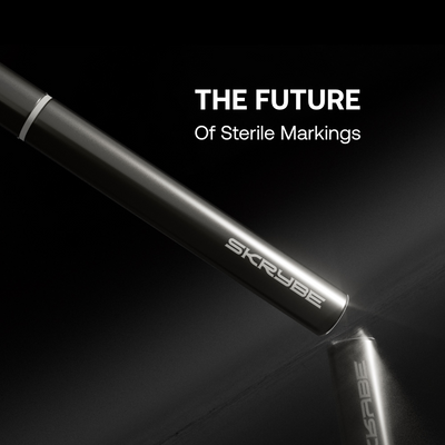
Visual mapping using sterile, skin-safe markers such as SKRYBE FMD ensures continuity between plan and practice, transforming the fold from a reactive treatment zone into a proactive structural opportunity.
The Anatomy of the Labiomental Complex
An accurate understanding of the anatomical layers of the labiomental area is essential. The fold itself is formed by the interplay between soft tissue structures and underlying skeletal anatomy, most notably the mandibular symphysis.
Key anatomical contributors include:
- Depressor labii inferioris (DLI): Inserts near the lower lip and contributes to dynamic lowering
- Mentalis muscle: Located just below the fold, it elevates the soft tissues of the chin and deepens the sulcus in action
- Orbicularis oris: Forms the sphincter muscle of the mouth, contributing to lip eversion and support
- Superficial fatty layer: Thins with age, exaggerating the shadowing effect of the fold
- Bony structure: The inclination and projection of the mandibular symphysis determines baseline fold depth

In youth, the labiomental angle typically ranges between 110–130 degrees, but this can deepen and become more acute (90–100 degrees) with bone resorption, fat loss, and soft tissue ptosis【1】.
Ageing and Morphological Variants
There are two primary presentations requiring intervention:
- Congenital/skeletal: Hypoplasia of the mandibular symphysis or vertical maxillary excess may result in deep folds even in younger patients.
- Acquired/ageing: Involutional changes including mentalis hyperactivity, soft tissue atrophy, and progressive mandibular recession deepen the sulcus and blunt the chin-lip transition.
Each variant demands a tailored mapping and treatment approach. A one-size-fits-all filler strategy risks either effacing necessary contours or accentuating disharmony.
Visual Planning and Marking Technique
Visual mapping is a critical step in managing the labiomental sulcus. When done post-cleansing, using sterile white-ink tools such as SKRYBE, it enables structural planning without compromising sterility or losing visibility during antiseptic application.
Mapping Process:
-
Midline Identification
Mark the vertical facial midline from the philtrum to the pogonion. This will guide symmetric volume placement. -
Sulcus Demarcation
Ask the patient to contract the mentalis. This will exaggerate the sulcus, allowing the clinician to trace its true depth and lateral extent—typically 2.5–3.5 cm across. -
Vector Lines
Mark shallow arcs inferiorly and slightly laterally from the mid-sulcus point. These vectors correspond to planned filler deposition paths, guiding cannula trajectory. -
Depth Planning
Mark anticipated injection planes with dot-notation—e.g. deep subcutaneous centrally, more superficial at the periphery to taper volume.
Markings should be symmetrical, photographically documented, and preserved throughout the procedure to support intraoperative orientation and post-procedure education.
Injection Technique
Injection strategy varies based on fold morphology and patient anatomy. However, certain principles remain consistent:
Cannula-Based Approach (Preferred)
- Cannula: 25G, 38 mm
- Entry Point: 1–1.5 cm lateral to sulcus, in line with mentolabial groove
- Plane: Deep subcutaneous to supraperiosteal
- Motion: Retrograde fanning or linear threading
- Volume: 0.3–0.6 mL per side, titrated to response

Needle Bolus Approach (Advanced Use)
- Reserved for deep skeletal deficits where bony projection is required
- Injected in controlled supraperiosteal boluses, with strict aspiration and low volume per pass
Filler Selection
Choice of product is dictated by intended correction:
- Hyaluronic Acid (HA) Fillers: High G’ and cohesivity options (e.g., Restylane Defyne, Teosyal Ultra Deep) work well for structural lift
- Collagen Stimulation: Products like Radiesse or Sculptra may be used to induce dermal thickening over time, particularly in long-term rejuvenation plans【2】
- Polynucleotides and Skin Boosters: Useful adjuncts for improving texture and elasticity, but not standalone treatments in structural fold correction
Correct product-layer pairing is paramount: deep folds require structural HA in deep planes; superficial wrinkles may be addressed with lighter rheology agents.
Common Errors and Complications
Errors from Lack of Mapping:
- Asymmetry: Misaligned vectors or unequal volume
- Overcorrection: Flattened sulcus resulting in a “ballooned” perioral appearance
- Vascular compromise: Mental artery and inferior labial branches lie nearby
Aesthetic Complications:
- Lumpiness: Occurs when filler is misplaced in the dynamic superficial plane
- Step-off deformity: Created by abrupt transition between filled sulcus and untreated chin or lower lip
All of these are avoidable with adequate pre-injection marking and understanding of individual structural variables.
Integration into Clinical Workflow
A well-structured marking protocol not only guides the injector but supports patient education and enhances consent. The use of SKRYBE sterile markers, applied post-antisepsis, allows:
- Permanent visibility throughout the session
- Clear anatomical anchoring for planned vectors and depth
- Documentation support for before-and-after comparison
Such visual tools are particularly useful in teaching environments, where junior injectors can be trained to respect anatomical norms and boundary conditions.
Discussion
The labiomental fold is frequently overshadowed by the more glamourised aspects of perioral rejuvenation. Yet, its treatment demands a nuanced understanding of both static structure and dynamic expression.
Too often, filler placement in this zone is dictated by visual deficit alone, without proper respect for skeletal contribution, muscular activity, or vascular proximity. Mapping provides a tactile and visual rehearsal of the procedure—anticipating issues before they arise.
In a systematic review by Park et al. (2022), the absence of structural planning was associated with significantly higher rates of re-treatment and patient dissatisfaction in labiomental correction procedures【3】.
Incorporating sterile, resistant marking tools like SKRYBE into procedural protocol not only improves safety but also communicates professionalism, hygiene, and intentionality.
Conclusion
Chin-sulcus correction is a structurally important but technically sensitive intervention in facial rejuvenation. Through comprehensive anatomical understanding, strategic visual mapping, and sterile marking implementation, clinicians can elevate outcomes from reactive correction to planned harmony.
Marking is not a preliminary sketch—it is the architectural blueprint. In the labiomental zone, where millimetres matter, that blueprint is indispensable.
References
- Rohrich, R. J., Pessa, J. E. (2007). The youthful lip: effects of aging on the upper lip and perioral complex. Plastic and Reconstructive Surgery, 120(5), 1041–1047.
- Sundaram, H., & Carruthers, J. (2015). Evaluation of the safety of calcium hydroxylapatite for soft-tissue augmentation. Plastic and Reconstructive Surgery, 136(5 Suppl), 81S–90S.
- Park, K. Y., Kim, B. J., & Kim, M. N. (2022). Efficacy and satisfaction of labiomental fold augmentation with filler injection: A systematic review. Journal of Cosmetic Dermatology, 21(3), 1021–1028.
- Coleman, S. R., & Grover, R. (2006). The anatomy of the aging face: volume loss and changes in 3-dimensional topography. Aesthetic Surgery Journal, 26(1S), S4–S9.
- Kang, S. H., & Hwang, K. (2018). The location and course of the mental artery in the labiomental fold. Annals of Plastic Surgery, 80(5), 524–528.


